MIPAR
  • Products
    • Analysis Software
    • Microscope Software
    • Compliance Software
    • Deep Learning
    • APIs
    • MIPAR for Academics
    • Request a Quote
  • Applications
    • Materials
    • Life Science
    • Manufacturing
    • BioMedical
    • Drone & Survey
    • Recipe Templates
    • Articles & Citations
    • Webinars
  • Spotlight ✧˙˖
  • User Resources
    • Udemy Course
    • Help desk
    • Forum
    • Tutorials
    • FAQ
    • User Manual
  • About
    • Who we are
    • Data Security
    • Newsletter
  • Download Trial
  • Home
  • > 
  • Applications
  •  > 
  • Life
  •  > 
  • Histology
  •  > 
  • Pathology

Digital Pathology Image Analysis with MIPAR

Precision and Automation for Histopathology
MIPAR is revolutionizing digital pathology image analysis with its automated software, offering both fully automated and supervised workflows. Our sophisticated software suite is specifically designed to enhance the efficiency and precision of histopathology research, transcending traditional manual microscopy methods. Simply open your pathology research images in MIPAR, apply your custom analysis protocol, and swiftly generate comprehensive, accurate reports.
 
Comprehensive Digital Pathology Image Analysis Overview
Digital pathology image analysis is integral to the study of diseases at a cellular and molecular level. It involves examining tissue samples and cellular structures to understand disease mechanisms, progression, and potential therapeutic targets. This analysis is fundamental in advancing medical knowledge and developing new treatments in the field of histopathology.
 
Overcoming Challenges in Histopathology
Manual pathology analysis in research settings can be time-consuming and subject to variability, especially when dealing with complex tissue structures or various disease states. Automated solutions are often needed to handle the vast array of data and intricate details present in digital pathology image analysis. MIPAR addresses these challenges with precision and efficiency.
 
MIPAR’s Solution for Digital Pathology Image Analysis
MIPAR tackles these challenges by integrating state-of-the-art image processing algorithms with advanced Deep Learning technology. Our software is adept at accurately identifying and quantifying pathological features such as cellular anomalies, tissue disorganization, and biomarker expression. MIPAR enables comprehensive tissue analysis, providing valuable insights for histopathology research.
 
Advantages of Using MIPAR for Digital Pathology Image Analysis
Implementing MIPAR for digital pathology image analysis results in significant time savings, enhanced consistency, and greater depth in data analysis. It provides a robust platform for detailed examination of pathological samples, contributing to the advancement of medical research and histopathology.
 
Who Can Benefit?
MIPAR is perfect for pathology researchers, scientists, and laboratory professionals who require precise, efficient, and comprehensive digital pathology image analysis tools for their histopathology research studies.
 
Technical Details
MIPAR supports a variety of image formats from different types of microscopy used in digital pathology image analysis. The software includes features for automated calibration, metadata import, and batch processing capabilities. Operating on local workstations, MIPAR provides a secure and effective analysis environment.

System Requirements
Documentation >>

Demo
See MIPAR's capabilities in pathology research in action. Schedule a demonstration with one of our Applications Engineers to discover how our software can transform your research endeavors.

Support
Our Applications Engineers, with their deep expertise in pathology and automated image analysis, are committed to supporting your research projects. Our support team provides comprehensive guidance to ensure you maximize the benefits of MIPAR's capabilities.

Cost
Licensing costs vary based on the type of license and the number of users. For specific pricing information tailored to your pathology research needs, please submit a quote request, and a representative will respond promptly.

QUOTE
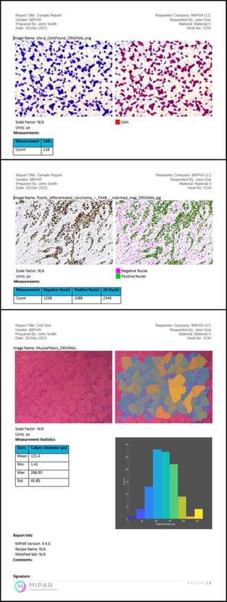
Sample Aluminum Grain Size Report

Resources
​​
•  Citations
​•  Benefits of All-Digital Histology
​•  What is Digital Pathology?

Picture

Check Out Our Products

Picture

MIPAR Base: our core product to analyze images from almost any source. Extensions enable Deep Learning models, Word reports and 3D data analysis.  MIPAR Base Page >>

Picture

​Microscope Software: capture, analyze and report all in a single workflow with MIPAR Live. Check to see if your camera is supported. MIPAR Live Page >> ​​

Picture

Compliance Software: analyze, report, and approve in a 21 CFR Part 11 or GMP Annex 11 compliant environment with traceability. MIPAR Checkpoint Page >> ​​

Picture

MIPAR APIs: tools to help integrate MIPAR detection and measurement solutions into other software applications and workflows. APIs Page >> ​​​

Picture

Call

Give us a call
​Mon-Fri 9am-6pm EST​​
​​+1-614-407-4510

Message

Tell us how we can help!

Chat

Chat with us
​Mon-Fri 9am-6pm EST​​
Chat with us ››

Home

Support

Contact

© 2025 |  Privacy  |  Terms
  • Products
    • Analysis Software
    • Microscope Software
    • Compliance Software
    • Deep Learning
    • APIs
    • MIPAR for Academics
    • Request a Quote
  • Applications
    • Materials
    • Life Science
    • Manufacturing
    • BioMedical
    • Drone & Survey
    • Recipe Templates
    • Articles & Citations
    • Webinars
  • Spotlight ✧˙˖
  • User Resources
    • Udemy Course
    • Help desk
    • Forum
    • Tutorials
    • FAQ
    • User Manual
  • About
    • Who we are
    • Data Security
    • Newsletter
  • Download Trial