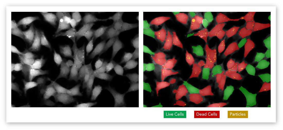
The Basic Principles of Cell Identification
All living organisms are composed of cells, from unicellular protozoa to the largest cetaceans in the world’s oceans. The simple fact that even the human body is reducible to tens of trillions of structural units is one of the core tenets of cell theory; now universally accepted as fact. This pioneering idea laid the groundwork for modern cellular biology and a plethora of discoveries since.
Biotechnology, molecular genetics, vaccinations, and more; none would be possible if not for our ability to characterize structures and dynamics at the cellular level. The history of cell identification is subsequently tied to that of microscopy.
Brief History of Cell Identification
Resolving power is a critical metric when it comes to cell identification and life sciences. This defines the ability to optically distinguish adjacent objects that are close together.
The average human eye has a resolving power approaching 200 micrometers (μm), which makes it impossible to observe even the largest human cell – the ovum (approximately 10 μm) – with the naked eye. It was not until renowned scientist Robert Hooke built a novel optical system based on existing compound microscopes that we could achieve magnifications great enough to visualize the microscopic world. Today, advanced compound microscopes have reached the limits of visible light with a maximum resolving power of approximately 0.2 μm, or 200 nanometers (nm).
However, cells are not only small but extremely complex. Even with the greatest of magnifications, it can be difficult to uncover their molecular composition and discover their functions based purely on light-based microscopy and visual cell identification. For that, biochemists largely rely on fluorescent tagging with tried-and-tested fluorophores like green fluorescent protein (GFP), propidium iodide (PI), and acridine orange (AO). These unique reagents bind to cells depending on their composition, viability, and so on, emitting characteristic fluorescent signals when excited by light in a given wavelength range. This makes it easier to visually count and identify cells based on a wide range of physicochemical properties.
Challenges of Cell Identification
There are many difficulties associated with modern cell identification, but one of the most pervasive is the margin for human error. The sheer abundance of cells in a sample coupled with the element of subjectivity when it comes to visual characterization means it can still be difficult to accurately identify cells in solution with the level of quantitative assurance that modern life sciences demand.
Biochemistry facilities increasingly rely on automated imaging systems to distinguish the weak fluorescent signals emitted by assayed samples and to eliminate the margin for human error.
Cell Identification with MIPAR

